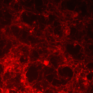
The application of an array of fluorochromes makes it possible to identify cells and cellular components with a high degree of specificity amidst non-fluorescing material. A fluorescence microscope is capable of revealing the presence of a single molecule, and through the use of multiple labeling, different probes can identify several target molecules simultaneously. The fluorescence microscope has the capability of detection of fluorescing molecules below the diffraction limit of specific specimen features.
Contact us to discuss your project!